Tricuspid Valve Surgery

What is the tricuspid valve?

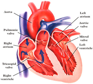
The heart consists of four heart valves. Two of them are on the left side and the other two are on the right side. The tricuspid valve is the inlet valve of the right side of the heart and is located between the two chambers. The tricuspid valve allows unobstructed flow of blood into the pumping chamber (right ventricle) of the heart without any resistance. 

Unlike the mitral valve, the tricuspid valve has three triangulated doors (leaflets) on the frame(annulus) and is held in place with strings allowing doors to open only one way ensuring blood flow only in a forward direction. If the strings break or get loose,  or the frame gets bigger,this leads to formation of gaps between the doors which causes leakage or backward flow. This can also occur if the doors get damaged due to infection. 

When the tricuspid valve becomes diseased, infected or malfunctions, it needs to be repaired or replaced with an artificial heart valve or other available alternatives.


Types of tricuspid valve disease

It is quite uncommon for the tricuspid valve to become faulty on its own unless the patient has developed severe infection. Most commonly, tricuspid valve disease is the end result of the other valve diseases or lung diseases. 

Tricuspid valve malfunction can result from mainly two mechanisms

  1. Tricuspid  Stenosis – In this disease, the opening of the tricuspid valve is restricted due to structural problems with the valve or surrounding tissues. Typically, the common causes for this condition are secondary to infection like rheumatic fever, or rare tumours.


  1. Tricuspid regurgitation – in this disease, the tricuspid valve does not close effectively leading to blood flowing backwards(leakage). The common cause for this type is due to outward stretch on the frame(annulus) of the tricuspid valve which  causes a gap in the center of the valve causing it to leak.  The other causes are bacterial infections(endocarditis), long standing atrial fibrillation, permanent pacemaker and a weak, enlarged heart. 


When is the valve repaired or replaced? 

Repairability of the valve depends on the causes of leakage, damage to the valve, complexity of surgery , patient choice and experience of the surgeon. In general, valve leakage secondary to a stretched frame is  readily repairable in the hands of most surgeons, with very good long term results. If the leakage is secondary to bacterial infection then repairability depends on the damage caused by infection and experience of the treating surgeon. Problems due to rheumatic fever are difficult to repair and such repairs are undertaken by highly specialist surgeons.The majority of surgeons will elect to replace tricuspid valves diseased by rheumatic fever. Leakage due to long standing atrial fibrillation is readily repairable in most cases as the cause of leak is the stretched frame.  Whatever the case may be, your surgeon will assess the valve after the repair and if not satisfied , your valve will be replaced with what you would have chosen prior to surgery. 

Is valve repair better than replacement?

Valve repair is generally considered better than replacement. This is due to the fact that you retain your tissue which is resistant to infection, compared to an artificial heart valve. An artificial heart valve is a large foreign body implanted into your circulatory system which is in constant contact with blood. There is a risk of  blood clot formation,  which can cause artificial valves to fail. If a patient gets any blood infection, then there is a risk of an artificial heart valve getting infected, which requires major surgery to replace the valve. Both the risks above are significantly lower with valve repair. Valve repair also requires the use of artificial material but the quantity of that is much lower compared to artificial heart valves. 

The other issue is the requirement of blood thinners like warfarin with artificial mechanical/metal valves. If you get a metal heart valve implantation then you need to be on warfarin for life, which is a strong blood thinner. It exposes patients to a life long risk of bleeding. With valve repair, patients do not need to take warfarin for life. 

If a patient gets a tricuspid valve replacement with a tissue valve then he/she may not need to take warfarin. It is thought that tissue valves may last longer in the tricuspid position compared to other heart valves, but data is not robust. Nonetheless, tissue valves have a good record in the tricuspid position. Younger patients are very likely to outlive tissue valves which will definitely require reoperation or a procedure in future. Unlike this, a valve repair can last for many decades in the majority of patients. 

Surgery for tricuspid valve repair:

The fundamental principle is to retain the patient’s own tissues and correct the faulty mechanism to make the valve function properly again. Valve repair requires supporting the valve with artificial strings, or removing the small diseased portion of the valve. After one of these is done, a semi circular ( shape of valve) ring is implanted outside the valve to hold the valve in its natural shape. Depending on the extent of the problem with the valve, your surgeon may need to resort to a few other techniques to repair your valve. As mentioned earlier, tricuspid valve disease is secondary to other heart issues. Your surgeon will need to correct the primary problems of the heart with appropriate surgery.  After the repair is done , the heart is started again and separated from the heart lung machine . Valve function is checked with (TOE)transesophageal echocardiogram( echocardiogram performed via oesophagus). Surgery then proceeds as routine if the surgeon and anaesthetist are happy with the valve performance. In some instances, the heart will be stopped again to do more touch ups to the repair to improve its function and in some cases, surgery will proceed to replace the valve if the valve is deemed not repairable after a few attempts. 


Surgery for tricuspid valve replacement:

This is typically done when the valve is deemed not repairable. Types of prosthesis will be discussed at the time of consultation and the prosthesis of your choice will be implanted. In rare instances, surgeons may override your choice to ensure your safety. 

Part of the valve may be excised if destroyed by infection, while some parts of its attachment to the heart are retained for better functioning of your heart. Artificial heart valves are implanted using multiple sutures and tied in place. Once the surgeon is happy with the mechanics of the valve, surgery will proceed as usual. The heart will be separated from the heart lung machine.  Valve function is again checked with (TOE) transesophageal echo and it is ensured that there are no major leaks. Small leaks may be accepted. Surgery then proceeds as routine if the surgeon and anaesthetist are happy with valve performance.


Do all heart surgeons do this operation?

All heart surgeons should be able to do this surgery, but some are more comfortable than others. 


Approaches to tricuspid valve surgery:

There are different approaches to carry out tricuspid valve surgery 

  • Sternotomy – standard approach. Involves splitting of the entire breastbone(sternum). The majority of patients who require tricuspid valve surgery also need other heart surgeries for which sternotomy would be necessary anyway.
  • Minimally invasive approach – involves 5-7 cm incision on the right chest and the valve is repaired by operating via the space in between the ribs
  • Tricuspid clip – performed via a little puncture in the groin. It is the inferior option compared to the above approaches in patients who are suitable for surgery. Only reserved for patients who are not suitable for surgery as the leakage can only be minimised instead of eliminating it. It cannot be used for a tricuspid valve with narrow orifice (mitral stenosis)


Surgery via standard sternotomy:

Surgery is done under general anaesthesia. It can take anywhere between 3 to 4 hrs. Surgery is performed after splitting the breast bone with a midline incision. The heart is connected to the heart lung machine and stopped while the heart muscle is protected with a special solution. 

Dr Joshi will open the chamber of heart that houses the tricuspid valve. Depending on what is required, the tricuspid valve will be repaired or replaced with a prosthesis of your choice. For repair, Dr Joshi will implant a tricuspid ring which sits on the frame of the valve ensuring correction of shape and reducing leakage. If additional procedures are required then they are performed simultaneously eg. Ablation of atrial fibrillation, mitral valve surgery or aortic valve replacement etc.  After finishing the surgery on the heart, it will be separated from the heart lung machine. Heart valve function will be checked by intraoperative echocardiography prior to returning the patient to ICU. The  ICU stay is generally 2 days and hospital stay is about 7 days. 

Typically most patients are kept asleep with medications for 3-6 hrs to observe for stability. While you are asleep you will be breathing with a breathing machine. The ICU specialist (Intensivist) is in charge of your care while you recover in the ICU. When you wake up you will feel very sleepy and weak. You will notice a few lines , tubes and wires connected to you which are routine. You will feel a bit uncomfortable with all those attached to your body. These are removed as you progress in your care and most are removed by day 5. As a routine, you will be helped to get out of bed and sit in a chair for a few hours to help your lungs breathe better. The physiotherapist will take you for a short walk on day 2 in the ICU. Stay in ICU is typically for 2 days. You will be transferred to the cardiac ward for further care.

Pain is very subjective but overall its uncommon for patients to experience severe pain. You will be given strong pain killers some of which may have unpleasant side effects like nausea, vomiting and constipation. Nursing staff will manage your pain medication in close collaboration with you. You are encouraged to notify staff if you your pain is unbearable, prevents you from deep breathing or keeping you awake.

Pain is very subjective but overall it is uncommon for patients to experience severe pain. You will be given strong painkillers, some of which may have unpleasant side effects like nausea, vomiting and constipation. Nursing staff will manage your pain medication in close collaboration with you. You are encouraged to notify staff if your pain is unbearable, prevents you from deep breathing, or keeps you awake. 


Progress in the ward

You will be in a room once you arrive on the ward. You will be connected to a monitor so nurses can see any alarming signals from your heart rhythm. You need to be careful that the monitor leads remain attached to your body properly. If they get detached, you should inform your nurse by pressing the call button. The most important thing in the ward is to start getting more mobile. You will be asked to walk 5 times every day and your physio will teach you exercises. Dr Joshi will give instructions for removal of pacing wires. Removal of wires and lines are very tolerable. By day 5-7, you will be asked by the physiotherapist to complete two flights of stairs. This is done to assess your fitness to be discharged home and your independent mobility. You will have regular blood tests and chest x rays. Electrolytes are frequently replaced with intravenous infusions. 


Going home

Most patients are expected to be discharged between 7-10 days after surgery. Staying after this type of surgery is usually longer compared to other heart surgeries. You will receive discharge education from your nurse who will give you information about incisions, dressings, showering etc. A pharmacist will visit you to discuss your medications which might have changed a little bit. You must remember to take your discharge file with you.This will include all your instructions regarding follow up appointments, a medication list and other material. You should carry this file with you when you visit your cardiologist or cardiac surgeon for follow up at 4-6 weeks

Dressings

You will have a long dressing on your chest. You should remove it after 4 weeks under a running shower. You can remove it earlier if it starts to come off or breaks. If you have any concerns about wound please visit your GP or notify Dr Joshi


Complications:

The risks of surgery can vary quite a bit and vary according to patients. As mentioned, tricuspid valve disease is a manifestation of other long standing heart diseases and the risks may be elevated compared to other surgeries. Not uncommonly, quite a few other heart surgeries are required along with tricuspid valve surgeries and the elevated risk is a combination of multiple factors that include delayed presentation, multiple operations and condition of the heart. The most important complications to keep in mind are the risks of bleeding and stroke. Post operative bleeding is due to the long operation and disturbance in blood clotting mechanisms after surgery. The specific risks are discussed at the time of surgery which also include infection and kidney failure 

One of the important non major complications with this surgery is a risk of permanent pacemaker (PPM), which is about 5%. The valve is placed immediately next to your heart’s electrical system which may get compressed and malfunction. If your electrical system does not recover within a week then Dr Joshi will ask the cardiologist to insert a PPM under local anaesthetic prior to your discharge from hospital. Insertion of PPM does not hold you long in hospital or affect your recovery adversely.

Younger patients are a risk of developing fluid build up around the heart. This typically manifests at about 4 weeks. This is reactive fluid and younger patients are more prone. It is not harmful but can make some patients feel short of breath or feverish. If that happens you should present to ED or notify your cardiologist or Dr Joshi. The treatment involves anti-inflammatory medications and in some cases taking the fluid out with a needle or small incision. 

If the risks of surgery are elevated then you will be informed about it at the time of consultation. 

Duration and type of blood thinner depends on the type of valve that is implanted in the heart. If you have a tissue valve implantation then you will be on warfarin for 3 months and if you have a mechanical/metal valve then you will be on warfarin for life. When surgery is elective, this will be discussed with you prior to surgery. 

If you are fitted with a tissue valve then Dr Joshi or your cardiologist will advise when you are able to stop warfarin. Generally, warfarin is replaced with Aspirin 100 mg daily after 3 months.

If you have a valve repair then your warfarin will be stopped after 6-8 weeks and replaced with Aspirin 100 mg daily.

Dr Joshi will see you for follow up after 6 weeks and then your long term follow up is organised by your cardiologist and GP. Echocardiogram is done once or twice a year to monitor the function of your heart and is done as seen necessary by your cardiologist. If you are on warfarin then your GP will guide you regarding frequency of blood tests and dosage of warfarin.

One of the reasons to repair or replace your valve is to enable you to return to your normal life. If you have any limitations due to your heart issues then you should also get better, effectively improving the quality of your life. One would be expected to return completely back to normal life. However, if you are on warfarin or other blood thinners then necessary changes to your lifestyle are recommended.

It is advised that you will need antibiotic prophylaxis for any future invasive procedure. You need to disclose to a treating practitioner or dentist about artificial prosthesis in your heart.