Mitral Valve Surgery

Minimally invasive mitral valve repair

Different approaches for mitral valve repair/replacement

  1. Sternotomy – splitting the breast bone
  2. Minithoracotomy – minimally invasive approach via small incision on the right chest

Minimally invasive mitral valve surgery

This approach involves a 5-7 cm incision on the middle side of the right chest. There is another small incision in the right or left groin. The surgeon approaches the mitral valve via the space in between the ribs. The ribs are spread with the insertion of a retractor allowing the surgeon to have improved exposure of the heart. Another 5 mm incision is placed on the top of the chest to introduce a telescopic camera that assists the surgeon’s visualisation of the heart and mitral valve.

minimally-invasive-heart-surgery

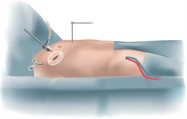
Incision in the groin is used to insert tubes in the heart to connect to the heart lung machine. The heart is stopped with a special solution. Valve is repaired or replaced depending on its condition. Heart is separated from the heart lung machine and allowed to beat normally on its own. At this stage, the anaesthetist and surgeon will assess the valve with an intra operative echocardiography to assess the valve function. Once satisfied, surgery will proceed in the usual manner to finish. Patient is transferred to the ICU still under anaesthetic.

If there are unexpected findings or difficulties, Dr Joshi will change the incision to a standard sternotomy incision to ensure your safety and good outcome. A good outcome is more important than incision. The occurrence of this event is quite uncommon.

What happens in the ICU?

Typically most patients are kept asleep with medications for 3-6 hrs to observe for stability. While you are asleep you will be breathing with a breathing machine. The ICU specialist (Intensivist) is in charge of your care while you recover in the ICU. When you wake up you will feel very sleepy and weak. You will notice a few lines , tubes and wires connected to you which are routine. You will feel a bit uncomfortable with all those attached to your body. These are removed as you progress in your care and most are removed by day 5. As a routine, you will be helped to get out of bed and sit in a chair for a few hours to help your lungs breathe better. A physiotherapist will take you for a short walk on day 2 in the ICU. The stay in ICU is typically for 2 days. You will be transferred to the cardiac ward for further care.

Will I be in a lot of pain?

Pain is very subjective. Pain from the chest wall incision is usually more severe than the incision on the breast bone. You will be on strong painkillers, some of which may have unpleasant side effects like nausea, vomiting and constipation. Nursing staff will manage your pain medication in close collaboration with you. You are encouraged to notify staff if your pain is unbearable, prevents you from deep breathing or keeps you awake.

Some patients may develop long term nerve pain that may result from the injury to nerves under the ribs. This is one of the downsides of this approach. Some patients may get fractures in the ribs that may take 6-8 weeks to heal.

Progress in the ward

You will be in a room once you arrive in the ward. You will be connected to a monitor so nurses can see any alarming signals from your heart rhythm. You need to be careful that the wires remain attached to your body properly. If they get detached, you should inform your nurse by pressing the call button. The most important thing in the ward is to start getting more mobile. You will be asked to walk 5 times every day and your physio will teach you exercises. You may have some wires connected to you which will be removed in the next few days. Removal of wires and lines are very tolerable. By day 5-7, you will be asked by physiotherapists to complete two flights of stairs. This is done to assess your fitness to be discharged home and your independent mobility. You will have a regular blood test and chest x rays. Electrolytes are frequently replaced with intravenous infusions.

Going home

Most patients are expected to be discharged between 5-7 days after minimally invasive surgery. You will receive discharge education from your nurse who will give you information about incisions, dressing, showering etc. A pharmacist will visit you to discuss your medications which might have changed a little bit. You must remember to take the discharge file with you that should include all your instructions regarding follow up appointments, a medication list and other material. You should carry this file with you when you visit your cardiologist or cardiac surgeon for follow up at 4-6 weeks.

Dressings

You will have a small dressing on the chest and one on the groin. You should remove it after 4 weeks under a running shower. You can remove it earlier if it starts to come off or breaks. If you have any concerns about the wound please visit your GP or notify Dr Joshi.

Follow up

You should arrange an appointment to see Dr Joshi in 6-8 weeks and your cardiologist in 8 weeks. You should visit your GP within a week after being discharged from hospital.

Advantages of minimally invasive approach:

Some of the advantages of a minimally invasive approach is reduced blood loss, shorter hospital stay and earlier return to activity. The breastbone (sternum) is kept intact which allows early resumption of activities like lifting weight or driving.

  1. Cosmetic – incision is small and in some patients barely visible
  2. Early recovery and return to activities
  3. Future reoperations on heart are less complicated if you need one
  4. Less blood loss, shorter ICU and hospital stay

Complications:

Generally considered a low risk operation despite the complexity associated with it. In the absence of major medical problems most patients have 1% or less risk of death or major complications. The most important complications to keep in mind are the risk of bleeding and stroke. Post operative bleeding is due to the requirement of delicate suturing in the high pressure part of the heart, and stroke can result from any debris including blood clots blocking one of the vessels in the brain. The incidence of bleeding and stroke is expected to be around 1% and if the risks are higher it will be discussed at the time of consultation.

One of the important non major complications with this surgery is a risk of permanent pacemaker (PPM) which is about 1%. If your electrical system does not recover within a week then Dr Joshi will ask the cardiologist to insert a PPM under local anaesthetic prior to your discharge from hospital. Insertion of PPM does not hold you long in hospital or affect your recovery adversely. It is quite common to develop irregular heart beat (30%). This is treated with medications for good effect.

Younger patients are a risk of developing fluid build up around the heart. This typically manifests at about 4 weeks. This is reactive fluid and younger patients are more prone. It is not harmful but can make some patients feel short of breath or feverish. If that happens you should present to ED or notify your cardiologist or Dr Joshi. The treatment involves anti-inflammatory medications and in some cases taking the fluid out with a needle or small incision.

Chest wall hernia is one of the uncommon complications of this approach in which a part of the lung can protrude between your ribs at the site of incision. This is not a major complication but may need surgery in some patients.

Phrenic nerve injury is a very uncommon complication. The phrenic nerve is a nerve that supplies the breathing muscle on the right side of the lung. There is a possibility of its blood supply being damaged or injury to the nerve itself due to indirect traction during heart surgery.

Blood thinners (warfarin or aspirin)

The duration and type of blood thinner depends on the type of valve that is implanted or valve repair. If you have a tissue valve implantation then you will be on warfarin for 3 months and if you have a mechanical/metal valve then you will be on warfarin for life. When surgery is elective,this would be discussed prior to surgery.

If you are fitted with a tissue valve then Dr Joshi or your cardiologist will advise when you are able to stop warfarin. Generally, warfarin is replaced with Aspirin 100 mg daily after 3 months.

If you have a valve repair then you will be on warfarin for 6-8 weeks following which it will be replaced with Aspirin 100 mg daily.

What type of follow up after surgery?

Dr Joshi will see you for follow up after 6 weeks and then your long term follow up is organised by your cardiologist and GP. Echocardiogram is done once or twice a year to monitor the function of your heart and is done as seen necessary by your cardiologist. If you are on warfarin then your GP will guide you regarding frequency of blood tests and dosage of warfarin.

Will my life be normal after surgery?

One of the reasons to repair or replace your valve is to enable you to return to your normal life. If you have any limitations due to your heart issues then you should also feel better, effectively improving the quality of your life. One would be expected to return completely back to normal life. However, if you are on warfarin or other blood thinners then necessary changes to your lifestyle is recommended.

Antibiotic prophylaxis prior to invasive procedures:

It is advised that you will need antibiotic prophylaxis for any future invasive procedure. You need to disclose to your treating practitioner or dentist about the artificial prosthesis in your heart.

Typically most patients are kept asleep with medications for 3-6 hrs to observe for stability. While you are asleep you will be breathing with breathing machine. The ICU specialist (Intensivist) is in charge of your care while you recover in ICU. When you wake up you will feel very sleepy and weak. You will notice few lines , tubes and wires connected to you which is a routine. You will feel a bit uncomfortable with all those attached to your body. The additionals are removed as you progress in your care and most are removed by day 5. As a routine, you will be helped to get out of bed and sit in chair for few hours to help your lungs breath better. Physiotherapist will take you for a short walk on day 2 in ICU. Stay in ICU is typically for 2 days. You will be transferred to cardiac ward after for further care.

Pain is very subjective but overall its uncommon for patients to experience severe pain. You will be given strong pain killers some of which may have unpleasant side effects like nausea, vomiting and constipation. Nursing staff will manage your pain medication in close collaboration with you. You are encouraged to notify staff if you your pain is unbearable, prevents you from deep breathing or keeping you awake.

Progress in ward

You will be in a room once you arrive to ward. You will be connected to monitor so nurses can see any alarming signals from your heart rhythm. You need to be careful that the wires remain attached to you body properly. If they get detached, you should inform your nurse by pressing call button. Most important thing in ward is to start getting more mobile. You will be asked to walk 5 times every day and your physio will teach you exercises. You may have some wires connected to you which will be removed in next few days. Removal of wires and lines are very tolerable. By day 5-7, you will be asked by physiotherapists to complete climbing of two flights of stairs. This is done to assess your fitness to be discharge home and your independent mobility. You will have regular blood test and chest x rays. Electrolytes are frequently replaced with intravenous infusions.


Going home

Most patients are expected to be discharge between 5-7 days after surgery. You will receive discharge education from your nurse who will give you information about incisions, dressing, shower etc. Pharmacist will visit you to discuss your medications which might have changed a little bit. You must remember to take discharge file with you that should include all your instructions regarding follow up appointments, medication list and other material. You should carry this file with you when you visit your cardiologist or cardiac surgeon for follow up at 4-6 weeks

Dressings

You will have a long dressing on chest. You should remove it after 4 weeks under running shower. You can remove it earlier  if it starts to come off or breaks. If you have any concerns about wound please visit your GP or notify Dr Joshi

Follow up

you should arrange an appointment to see Dr Joshi in 6-8 weeks and your cardiologist in 8 weeks. You should visit your GP withing a week after being discharged from hospital


Complications:

Generally considered a low risk operation despite complexity associated with it. In the absence of major medical problems most patients have 1%  or less risk of death or major complications. Most important complications to keep in mind is risk of bleeding and stroke. Post operative bleeding is due to requirement of delicate suturing in high pressured part of the heart and stroke can result from any debris including blood clots blocking one the vessels in the brain. The incidence of bleeding and stroke is expected to be around 1% and if the risks are higher it will be pointed out at the time of consultation.

One of the important non major complication with this surgery is a risk of permanent pacemaker (PPM) which is about 5%.The valve is placed immediately next to your heart’s electrical system which may get compressed and malfunction. If your electrical system does not recover within a week than Mr Joshi will ask cardiologist to insert a PPM under local anaesthetic prior to your discharge from hospital. Insertion of PPM does not hold you long in hospital nor affects your recovery adversely.

Younger patients are a risk of developing  fluid build up around the heart. This typically manifests at about 4 weeks. This is reactive fluid and younger patients are more prone. It is not harmful but can make some patients feel short of breath or feverish. If that happens you should present to ED or notify your cardiologist or Mr Joshi. The treatment involves anti inflammatory medications and in some cases taking the fluid out with needle or small incision.

If the risks of surgery are elevated then you will be informed about it at the time of consultation.

Duration and type of blood thinner depends on the type of valve that is implanted in heart. If you have a tissue valve implantation then you will be on warfarin for 3 months and if you have a mechanical/metal valve then you will be on warfarin for life long. This would be discussed with you prior to surgery when it is elective.

If you are fitted with tissue valve then Mr joshi or your cardiologist will advise when you are able to stop warfarin. Generally, warfarin is replaced with Aspirin 100 mg daily after 3 months.

If you had valve repair then your warfarin will be stopped after 6-8 weeks and replaced with Aspirin 100 mg daily.

Mr Joshi will see you for follow up after 6 weeks and then your long term follow up is organised by your cardiologist and GP. Echocardiogram is done once or twice a year to monitor the function of your heart and is done as seen necessary by your cardiologist. If you are on warfarin than your GP will guide you regarding frequency of blood test and dosage of warfarin.

One of the reasons to repair or replace your valve is to enable you to return to your normal life. If you had any limitations due to your heart issues then if should also get better, effectively improving quality of your life. One would be expected to return completely back to normal life. However, if you are on warfarin or other blood thinners then necessary changes to your life style is recommended.

It is advised that you will need antibiotic prophylaxis for any future invasive procedure. You need to disclose to treating practitioner or dentist about artificial prosthesis in your heart.