Atrial Septal Defect

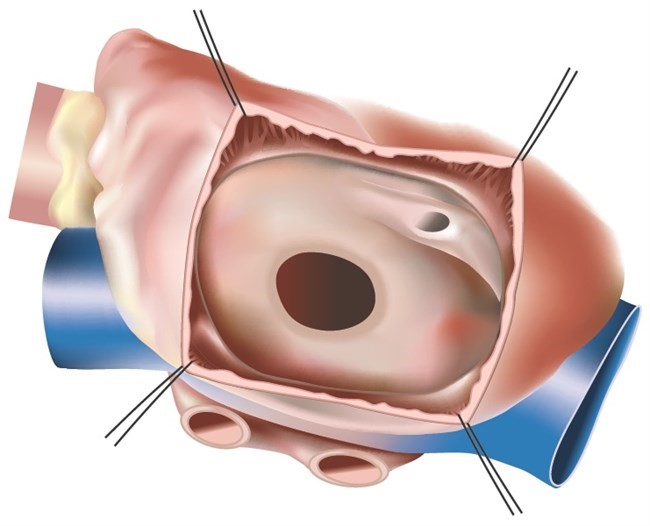
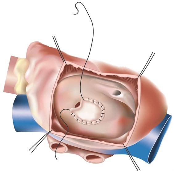
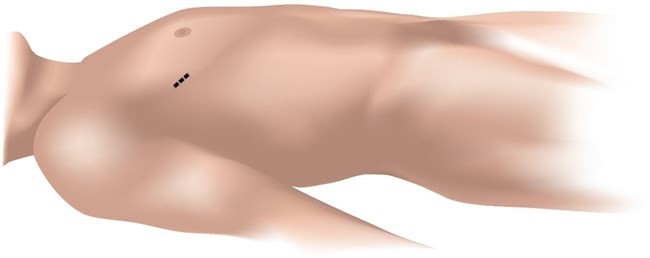
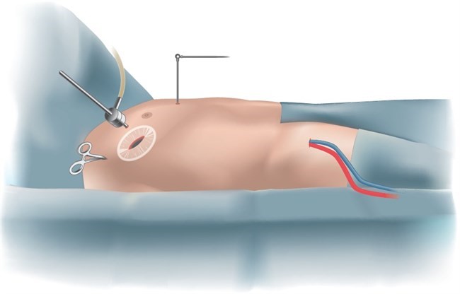
Minimally invasive closure of Atrial Septal Defect
An atrial septal defect (ASD) is a congenital heart defect where there is a hole in the atrial septum. The heart has two sides – right and left. The right side pumps unoxygenated blood to the lungs so blood can be oxygenated. The left side pumps oxygenated blood that it receives from lungs to the entire body. The two sides of the heart are separated by a tissue wall that ensures the blood does not mix between two sides. However, some people are born with defects in the wall allowing blood to flow between the two sides.

On each side of the heart there are two chambers. The chambers that collect blood are called the atrium and the chambers that pump blood out of the heart are called ventricles. The wall that separates the atrium is known as the atrial septum and the defect in this wall is called an Atrial Septal Defect(ASD). It can vary between 1 to 5 cm. We are all born with a small ASD at birth, but this usually seals off shortly thereafter. If the natural opening persists, it is called patent foramen ovale, the bigger opening is called ASD.

What are the symptoms of ASD?
ASD is quite often detected by accident. Symptoms typically include shortness of breath, fatigue, heart palpitations, or a decrease in exercise capacity. Patients usually have an audible heart murmur that is caused by the extra blood flow across the pulmonary valve to the lungs.

Is Is ASD potentially life threatening?
ASD causes excessive blood flow to the right side of the heart which can result in an enlarged right heart.This can then result in failure of the right heart and abnormal heart rhythms. Some patients can develop leakage of valves on the right side of the heart. The excessive blood flow in the right heart results in excessive blood flow in the lungs which in turn can damage vessels in the lung and causes life threatening complications like irreversible high pressures in lungs which can result in premature death or require heart lung transplantation.

When should an ASD be treated?
There are no rules but there is a general agreement that regardless of the age of the patient ASD should be treated. Some of the structural changes in the heart caused by an ASD over time are not entirely reversible. ASD closure before irreversible changes occur is therefore recommended. Most patients, children as well as adults, usually experience marked improvement of their symptoms after ASD closure.

How can an ASD be closed?

There are two ways to close the ASD.

  1. Percutaneous closure – when suitable, ASD can be closed by inserting a device that looks like a little circular mesh from a little puncture in the groin. It is done by a specialised cardiologist. Not all patients are suitable for this approach. Each patient is assessed and if suitable, will be offered device closure. The advantage of device closure is that one can avoid heart surgery and related recovery periods. The disadvantage is that the device can rarely dislodge which may require emergency heart surgery.
  2. Surgical closure – This involves open heart surgery.If the defect is small it can be closed with sutures,however if it is larger it will be repaired with a patch. Surgery can involve either a large incision on the chest or a small cut on the right side of the chest as a minimally invasive procedure. Only some surgeons specialise in the minimally invasive procedure.

Surgical Closure ASD
Surgical closure of ASD can be performed by two methods

  1. Standard sternotomy – This involves incision on the breastbone(sternum). The sternum is split in two halves to expose the heart.
  2. Minimally invasive approach – This does not involve splitting the sternum. Instead, it is performed by a small incision about 6 cm on the right side of the chest.

What is the difference between a sternotomy approach and the minimally invasive approach ?
Minimally invasive approach avoids splitting sternum hence sternum related complications such as infection, bleeding or malunion are avoided. Cosmetically, the minimally invasive approach is superior. Recovery is quicker with a minimally invasive approach and the limitations placed on activity are also fewer.

How is minimally invasive surgical closure of ASD performed ?
Surgery is performed under general anaesthesia. After preparation for surgery, Dr Joshi places an incision between 5-7 cm under the right breast(female)/right nipple(male). This allows the entry to the chest and exposure of the heart. An additional hole is made to introduce a camera system to visualise the heart. Surgery requires support of a heart lung machine which is connected by a small incision in the right groin. The blood vessels in the right groin are used to connect the heart lung machine. As mentioned earlier, the defect is in the partition that separates the right and left atrium. The defect is approached by opening the right atrium. Small defects can be sutured closed while the large defects are patched using bovine tissue. Right atrium is closed and the heart is separated from the heart lung machine. All incisions are closed and patient is transferred to ICU for further care

Are all patients suitable for minimally invasive surgery?
Most patients with an ASD are candidates for minimally invasive surgery however, patients with excessive weight or breast implant may not be considered for this approach.

Figure 1-Open ASD

Figure 2-Closed ASD

Figure 3-Incision

Figure 4-Surgical Setup

Patient is transferred to the ICU, still connected to the respirator. This is done purposefully to allow for the body to recover from surgery. After a few hours the patient is completely awake. There will be some drains connected to the chest wall and afew other monitoring lines. It feels awkward to be connected to wires and pipes but those are removed as you progress in your care. ICU stay is generally 1-2 days and hospital stay is about 5-7 days. Once in ward, active mobilization is recommended. This will be done with nursing staff and physiotherapists. Once recovered adequately, patients are encouraged to mobilize 5-6 times daily. While in the ward, blood tests are done regularly to check kidney functions and electrolytes. Echocardiogram, chest x ray and ECG are done prior to your discharge home.

Complications such as death, stroke , kidney failure are very uncommon and are expected to be less than 1%. There is always a risk of bleeding after surgery which is also 1%. Bleeding may be treated with blood products and having a second look operation to assess. The second look operation is usually short and does not have major side effects.

During your recovery you may develop an irregular heartbeat(AF) which is quite common. It is treated with medications with very good results. Side effects of anaesthesia and pain medications may be experienced such as nausea, vomiting, constipation, disturbed sleep, vivid dreams , depression or anxiety.

With a minimally invasive approach, some patients can get long term pain issues due to damage to nerves under ribs. They may need medications to control pain.

Generally complication rate is quite low and most patients recover well unless patients have risk factors prior to surgery.

It depends on individual circumstances and fitness prior to surgery but it is expected to recover near normal by 4 weeks after surgery. You should not drive for 4 weeks after your surgery. One is expected to completely return back to normal life as before.

You will be discharged with pain medications and medications that you were on prior to surgery. If patch is used for repair of the defect then you may be commenced on warfarin for 6 weeks followed by Aspirin.

You need to see Dr Joshi after 6 weeks for post operative assessment of your wounds and chest x-ray. You should arrange to see your GP after 1 week from discharge and your cardiologist after 4 weeks.

ASD doesn’t recur but the patch can come loose or sutures to repair ASD can fail which can lead to recurrence. This is very rare.

Closure of atrial septal defect is not associated with any long term side effects. One may develop side effects of the operation such as long term pain or effects related to scar.