Thoracoscopic occlusion of left atrial appendage with Atriclip

thoracoscopic-occlusion-of-left-atrial-appendage-with-atriclip

What is the left atrial appendage

Left atrial appendage is a small pouch on the left upper chamber of the heart. There is a similar pouch on the right upper chamber of heart. The main function of the pouch is to secrete a hormone that assists us in managing fluid balance in the body. Some believe that it also acts like a shock absorber if the chamber pressure rises.

How does the left atrial appendage cause stroke in patients with atrial fibrillation ?

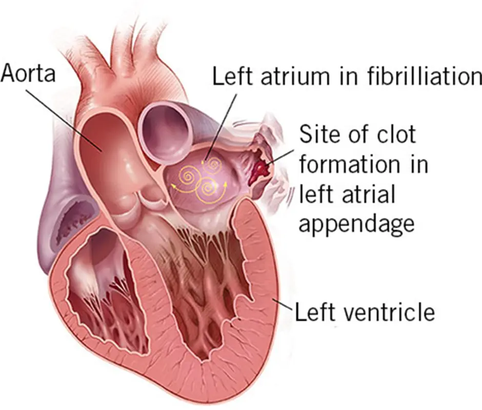
In a normal heart rhythm, the left atrium (collecting chamber) contracts/pumps blood out of the atrium into the ventricle(pumping chamber). The left sided chambers pump blood to the entire body including the brain. Hence, blood is constantly moving through the heart chambers. In patients with the rhythm of atrial fibrillation, the left atrium stops pumping blood effectively leading to stasis of blood within the chamber. Blood passively flows through the heart in patients with atrial fibrillation. One of the drawbacks of blood stasis in the left atrial appendage is that it can form a blood clot. If the clot/thrombus escapes from the left atrial appendage it will travel into the left pumping chamber (ventricle) and the pumping chambers pumps the clot out of the heart into the body. If the clot lodges into the brain arteries it can result in a major stroke. If it lodges into arteries of any other organ it can cause detrimental effects.

How can one prevent clot/thrombus formation in left atrial appendage and prevent stroke ?

The most common way to prevent thrombus/clot formation is to start patients on stronger blood thinners that include warfarin. The newer, most common blood thinners are Xarelto, Eliquis and Pradaxa. Warfarin requires blood tests for monitoring while the newer agents do not require blood tests.Because of this,it is more convenient for patients and has become more popular. Blood thinners thin the blood and minimise the chances of clot formation in the appendage thus reducing the risk of stroke.

One of the side effects of blood thinners is excessive bleeding from minor injuries and in some patients life threatening bleeding episodes. Some patients can experience bleeding from their nose from blowing or bleeding from gums while brushing and these are considered minor bleeding episodes. Other patients can suffer from life threatening bleeding eg – bleeding from gut, urinary tract or brain. Being on blood thinners affects the lifestyle of active young people eg – sports, hiking etc.

Another way of stopping clot formation is to occlude the cavity of the left atrial appendage. As blood cannot enter the appendage after its occlusion, the clot formation in the appendage is prevented. This is achieved by placing a clip on the neck of the appendage and occluding the appendage. This method is a good alternative to blood thinners (anticoagulants)

Who is a candidate for thoracoscopic occlusion of left atrial appendage ?

  • Patients with contraindications for blood thinners
  • Patients who cannot take blood thinners reliably
  • Patients in whom other means of occluding the appendage is not suitable
  • Patients who are non compliant or do not want to take blood thinners
  • Patients who are undergoing AF surgery

What are the other options other than Atriclip?

The left atrial appendage can be occluded with a device that is known as a Watchman device. It is inserted by a small puncture in the groin vessel, by the cardiologist. The cardiologist makes a puncture inside the heart to cross from the right side to the left side of heart. With the help of x-ray, a device that looks like an umbrella is inserted into the opening of the left atrial appendage. This device is anchored into its position with small barbs present on the outer surface of the device. It is advised that patients remain on anticoagulation (blood thinners) for 3-6 months as there is a chance of forming a blood clot on the device itself in the absence of blood thinners.

What is the difference between Atriclip and Watchman?

Atriclip is an externally placed device which is not in direct contact with blood while the Watchman is an internally placed device which is in direct contact with blood. No anticoagulation is necessary after insertion of Atriclip from the device point of view however, it is advised that patients stay on anticoagulation after insertion of Watchman device, as the device is in direct contact with the blood stream. Atriclip once placed doesn’t get dislodged, while there is a small chance of the Watchman device becoming dislodged, requiring urgent open heart surgery. Atriclip requires thoracoscopic procedures which is performed by 5 mm holes x 3 on the left chest while Watchman is inserted with a single puncture in groin. Both procedures require anesthesia and transesophageal echocardiogram to confirm appendage occlusion.

What is Atriclip ?

Atriclip is made from titanium metal and covered with cloth layers which helps the clip to be incorporated by tissue growth around it. It is MRI compatible (less than 3 Tesla). It is mounted on the special handle which is removed after its application on the base of the appendage.

Who does the Atriclip procedure ?

The procedure is done by a specialist Cardiothoracic Surgeon who has expertise in videoscopic surgery. This involves operating by looking at the pictures generated by a telescope introduced in the patient’s left chest with a 5 mm cut.

Pre procedural tests:

You will need to have an echocardiogram prior to surgery and in some cases Dr Joshi may request a CT Scan of your chest. You will also need a blood test, ECG and chest x-ray which are organised prior to surgery. Most patients are admitted the night before surgery and you will meet your anaesthetist in the hospital after admission.

Thoracoscopic occlusion of left atrial appendage (Procedure):

The procedure is keyhole surgery that involves three small incisions on the left chest, and is carried out under general anaesthesia. Procedural time is about 20-25 mins and total time including anaesthesia is about 50 mins.

Once referred, the patient is evaluated by Dr Joshi for the procedure. The procedure is discussed in detail with the patient. Preoperative preparation and investigations are done including echocardiogram and blood tests. There may be a requirement for a CT scan of the chest.

Blood thinners are typically stopped 48 hrs prior to surgery.

The patient is admitted to hospital the night before or the day of the procedure. The procedure is performed under general anaesthesia. The cardiac anaesthetist inserts monitoring lines in the neck and forearm. A urinary catheter is not used routinely. After the patient is asleep, the anaesthetist performs a transoesophageal echo (TOE) to examine the heart and to see whether there are any clots/thrombus present in the heart. If there is a thrombus/clot in the heart the procedure will not be performed as there is a risk of stroke if any procedure is performed with the presence of thrombus in the heart. Once the anaesthetist has confirmed the absence of thrombus in the heart, surgery is started.

The patient is placed in the supine position. 2 x 5 mm incisions are placed on the left side of the chest and a 12mm incision is placed on the lower left chest. Telescope/Camera is introduced in the chest via one hole and other two holes are used for inserting the instruments and the Atriclip.

The heart is covered by a loose sac. An opening of approximately 4 cm is made in the sac to expose the left atrial appendage. Dr Joshi will place an appropriately sized clip externally at the opening of the appendage. At the same time, the anaesthetist is examining with the TOE to confirm the satisfactory clip placement. Once confirmed,all the instruments are removed and all three incisions are closed with sutures.

Procedural and post procedural complications:

Complications are uncommon however, worth knowing. The most relevant complications are

  1. Cardiac perforation – Any procedure done on the heart involves a risk of injury to the chambers. If it occurs then Dr Joshi may have to extend the incision to control the bleeding.
  2. Pericarditis – This is the terminology for inflammation of the sac that covers the heart. This can be secondary to rubbing of instruments on the surface or sometimes due to a bloodless left atrial appendage. We have seen this complication in about 10% of patients. It is treated with anti-inflammatory medications with good results. In some patients it may manifest as fluid build up around the heart.
  3. Stroke – very rare complication. We check the appendage to make sure there is no clot or thrombus in it. Rarely a small clot or thrombus can go unrecognised which can cause stroke.

Hospital stay and recovery:

Most patients will be discharged 2-3 days after surgery. Patients are expected to be fairly independent and are able to take care of themselves. Recovery period is about 2 weeks. It is advised that patients do not drive for 2 weeks from the date of discharge. All activities can be gradually resumed as tolerated. There are no specific restrictions except for general care of your incisions. You will be given a list of medications and a list of the appointments to be made for follow up.

Follow up:

You need to see Dr Joshi 4-6 weeks after your procedure. He may organise CT scan or echo to assess the adequacy of occlusion of appendage. You will also need to make an appointment to see your cardiologist in 4-6 weeks.

The procedure is done by specialist Cardiothoracic Surgeon who has expertise in videoscopic surgery that involved operating by looking at the pictures generated by telescope introduced in the patients left chest with 5 mm cut.

You will need to have an echocardiogram prior to surgery and in some case Dr Joshi may request CT Scan of your chest. You will also need blood test, ECG and chest x ray which are organised prior to surgery. Most patients are admitted night prior to surgery and you will meet your anaesthetist in the hospital after admission

The procedure is a key hole surgery that involves three small incisions on the left chest and carried out under general anaesthesia. Procedural time is about 20-25 mins and total time including anaesthesia is about 50 mins.

Once referred patient is evaluated by Dr Joshi for the procedure. Procedure is discussed in details with patient. Preoperative preparation and investigations are done which includes echocardiogram and blood tests. There may be a requirement of CT scan of chest.

Blood thinners are typically stopped 48 hrs prior to surgery.

Patient is admitted to hospital a night prior or the same day of procedure. Procedure is performed under general anaesthesia. Cardiac anaesthetist inserts monitoring lines in neck and fore-arm. Urinary catheter is not used routinely. After patient is asleep, anaesthesiologist performs transoesophageal echo (TOE) to examine the heart and to see whether there are any clots/thrombus present in the heart. If there is a thrombus/clot in the heart the procedure will not be performed as there is a risk of stroke if any procedure is performed with presence of thrombus in the heart. Once the anaesthesiologist has confirmed the absence of thrombus in heart surgery is started

Patient is placed in supine position.2 x  5 mm incisions are placed on the left side of  the chest and 12mm hole is placed on the lower left chest. Telescope/Camera is introduced in the  chest via one hole and other two holes are used for inserting instruments and Atriclip. Heart is covered by a loose sac. An opening of approximately 4 cm is made in the sac to expose the left atrial appendage. Dr Joshi will place an appropriate size clip externally at the opening of appendage. While this is being done, anaethesiologist is examining and confirms the satisfactory clip placement with TOE. All the instruments are removed and all three incisions are suture closed.

Complications are uncommon however, worth knowing. The most relevant complications are

  1. Cardiac perforation – Any procedure done on the heart involves risk of injury to the chambers. It it occurs then Dr Joshi may have to extend the incision to control the bleeding.
  2. Pericarditis – This is the terminology for inflammation of the sac that covers the heart. This can be secondary to rubbing of instruments on the surface or sometimes due to bloodless left atrial appendage. We have seen this complication in about 10% of patients. It is treated with anti inflammatory medications with good results. In some patient it may manifest as fluid build up around heart.
  3. Stroke – very rare complication. We check the appendage to make sure there is no clot or thrombus in it. Rarely a small clot or thrombus can go unrecognised which can cause stroke.

You need to see  Dr Joshi between 4-6 weeks. He may organise CT scan or echo to assess the adequacy of occlusion of appendage. You will also need to make an appointment to see your cardiologist in 4-6 weeks