Valve sparing aortic root replacement surgery(David’s Procedure or VSARR)

What does this operation involve ?
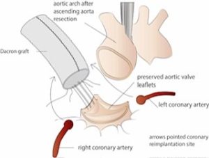
As the name suggests in this operation,aneurysmal Aortic root is replaced with sparing of the native aortic valve. In conventional aortic root replacement surgery, aortic root replacement includes replacement of the ascending aorta , aortic root and aortic valve with reimplantation of coronary arteries. In VSARR , everything is similar to conventional aortic root replacement, except that instead of an artificial valve,the patient’s own aortic valve is used.

What are the advantages of VSARR over conventional aortic root replacement ?
VSARR is very similar to conventional aortic root replacement except that the patient’s own aortic valve is retained instead of replacing it with a mechanical or tissue valve. All artificial valves expose patients to an additional lifetime risk of stroke and infection . Mechanical valves have an added lifetime risk of bleeding while tissue valves may require future interventions. Patient’s native valves have a lower risk of the complications associated with artificial valves.

Am I a candidate for VSARR ?
Not everyone is suitable for VSARR. The native valve should be in good condition and anatomically suitable for reimplantation. Some patients are born with two leaflets instead of three but they can still have this operation as long as their leaflets are in good condition. The risk of failure increases if it is done in patients who are not suitable for VSARR. The surgeons who are experienced (more than 30 operations in their career) will advise you whether you are a suitable candidate for VSARR. To assess your candidacy for this operation your surgeon will organise a CT scan and transoesophageal echocardiogram after assessing your initial investigations.

What are the indications of VSARR ?
Typically this operation is done in patients who have an aortic root aneurysm with relatively normal structure of the aortic valve. Very experienced surgeons also perform this operation in patients with an acute tear in the aortic root. This operation is not suitable for patients with aortic root aneurysm with calcified aortic valve leaflets.

What is the aortic root ?
The aorta is the main pipe or tube that originates from the outflow portion of the left side of the heart. Through this tube the heart pumps blood into the entire body. As the aorta goes out further from the heart it gives branches to various parts of the body to supply blood to all organs. Part of the aorta within the chest is known as the “ Thoracic aorta” and the part in the abdomen is known as “ abdominal aorta” and hence the term thoracic aortic aneurysm or abdominal aortic aneurysm. The diameter of this tube varies between 3.0 to 4.0 cm and it varies according to height, gender and race of the patient. There is an outlet valve at the beginning of this tube which is known as the “aortic valve”. This valve, when functioning normally, is a one way valve meaning blood can only go out of the heart and cannot re-enter the heart chamber.

Aortic root – this part, as the name implies, is the very origin of the aorta from the heart. The aortic root includes origin of aorta, aortic valve-which is an outlet valve and the origin of the coronary arteries that supplies blood to heart muscle.

Do all heart surgeons perform this surgery ?
This operation is done by specialist aortic root surgeons. Different heart surgeons have different types of specialist interest in heart surgery. This surgery is quite complex and also uncommon. Only a handful of surgeons are experienced enough to carry out this surgery routinely. The experience and expertise is limited by the limited number of patients in the community.

Surgery is done under general anaesthesia. It is a longish operation but depending on the simplicity or complexity it can take anywhere between 4 to 5 hrs. The heart is connected to a heart lung machine and stopped, while the heart muscle is being looked after and protected.

Dr Joshi will remove the aneurysmal part of the aortic root and ascending aorta, carving the tissues around the native aortic valve, leaving it attached to its skeleton. The left and right main coronary arteries are carefully separated. He then sutures a commercially available root( tube made from Polyester coated with gelatine) at the outlet of the heart and ties it down to ensure that the aortic root does not dilate in future. Next he sutures your native valve in this artificial tube in symmetric fashion. Depending on how well your valve is functioning, this may need to be repaired. Next, he will re-implant the right and left main coronary arteries back into their natural position to be able to supply blood to the heart muscle again. He performs a manoeuvre with a telescope (aortoscopy) to check the symmetry of your newly implanted valve. After finishing that, the heart is separated from the heart lung machine and takes over its job again. You will then return back to the ICU. ICU stay is generally 2 days and hospital stay is about 7 days.

It is generally considered a low risk operation despite the complexity associated with it. In the absence of major medical problems most patients have 1% risk of death or major complications. Most important complications, particularly with this operation to keep in mind, is risk of bleeding and heart attack. Post operative bleeding is due to the requirement of delicate suturing in the high pressured part of the heart, and heart attack if something goes wrong in re implantation of the left and right coronary arteries that are the main blood supply of the heart. The other complications are infection, kidney injury and lung related complications which are associated with any heart surgeries.

One of the important non major complications with this surgery is a risk of permanent pacemaker (PPM) which is about 5%. This surgery involves intricate dissection around the heart muscle close to the electrical system and there is a risk of accidental injury to it. If your electrical system does not recover within a week then Dr Joshi will ask a cardiologist to insert a PPM under local anaesthetic prior to your discharge from hospital. Insertion of PPM does not increase your stay in hospital or affect your recovery adversely.

Younger patients are a risk of developing fluid build up around the heart. This typically manifests at about 4 weeks. This is reactive fluid and younger patients are more prone. It is not harmful but can make some patients feel short of breath or feverish. If that happens you should present to ED or notify your cardiologist or Dr Joshi. The treatment involves anti-inflammatory medications and in some cases taking the fluid out with a needle or small incision.

You will be in a room once you arrive in the ward. You will be connected to a monitor so nurses can see any alarm signals from your heart rhythm. You need to be careful that the wires remain attached to your body . If they get detached, you should inform your nurse by pressing the call button. The most important thing in the ward is to start getting more mobile. You will be asked to walk 5 times every day and your physio will teach you exercises. You may have some wires connected to you which will be removed in the next few days. Removal of wires and lines are very tolerable. By day 5-7, you will be asked by physiotherapists to complete two flights of stairs. This is done to assess your fitness to be discharged home and your independent mobility. You will have regular blood tests and chest x rays. Electrolytes are frequently replaced with intravenous infusions.

Most patients are expected to be discharged after 7 days. You will receive discharge education from your nurse who will give you information about incisions, dressings, showering etc. A pharmacist will visit you to discuss your medications which might have changed a little bit. You must remember to take your discharge file with you, which will include all your instructions regarding follow up appointments,a medication list and other material. You should carry this file with you when you visit your cardiologist or cardiac surgeon for follow up at 4-6 weeks.

Dressings

You will have a dressing on your chest incision. You should remove it after 4 weeks under a running shower. You can remove it earlier if it starts to come off or breaks. If you have any concerns about the wound please visit your GP or notify Dr Joshi.

Follow up

You should arrange an appointment to see Dr Joshi in 6-8 weeks and your cardiologist in 8 weeks. You should visit your GP within a week after being discharged from hospital.

The advantage of this operation is that patients don’t require warfarin or stronger blood thinners. Due to placement of a polyester graft you will be on aspirin after surgery.

Dr Joshi will see you for follow up after 6 weeks and then your long term follow up is organised by your cardiologist and GP. Echocardiogram is done once or twice a year to monitor the function of your heart and is done as seen necessary by your cardiologist. If you are on warfarin then your GP will guide you regarding frequency of blood tests and dosage of warfarin.